The Beta Cell's Breakfast Audit: Tracing Every Step from Toast to Insulin
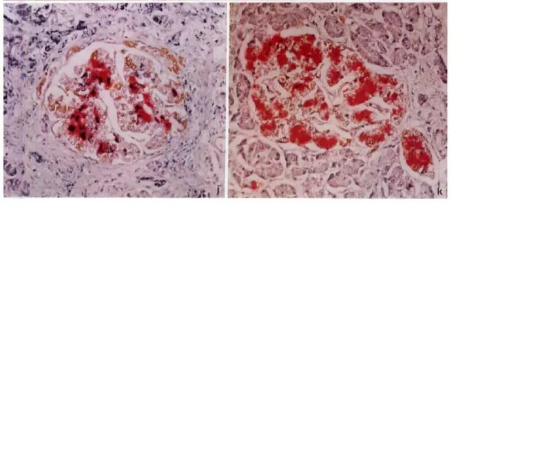
Photo: Hotheartdog, CC BY-SA 4.0, via Wikimedia Commons
Somewhere in your abdomen, roughly a million microscopic clusters of cells have just been handed a task. You have eaten — perhaps a full English, perhaps merely a morning biscuit with your tea — and the glucose from that meal is now arriving in your bloodstream. The clusters in question, the islets of Langerhans, must answer with insulin, and they must answer in the right amount, at the right moment. Get this calculation wrong too often, and the result is type 2 diabetes, a condition affecting nearly four million people in the UK.
How, at the level of individual molecules, does a beta cell read a breakfast? The answer is one of the most elegant signalling cascades in human physiology.
Glucose Enters Uninvited
The beta cell has a peculiar habit: it lets glucose flood in through a transporter called GLUT2 (in humans; rodents use GLUT2 and GLUT1 in differing proportions), without spending any energy on the process. Glucose does not knock. It simply follows its concentration gradient across the membrane.
Once inside, the sugar is phosphorylated by glucokinase — an enzyme with a deliberately low affinity for glucose. Glucokinase is often called the 'glucose sensor' of the beta cell, and the title is earned. Unlike its cousin hexokinase, which saturates quickly, glucokinase keeps working as glucose levels climb, meaning the cell's metabolic rate becomes a direct, faithful readout of blood sugar. The breakfast is, quite literally, being converted into metabolic current.
The Metabolic Trigger
Glucose now enters glycolysis and the mitochondrial citric acid cycle. The cell's mitochondria burn through the sugar, and two things happen that matter enormously for signalling.
First, the ratio of ATP to ADP inside the cell rises sharply. This is not merely fuel; it is the message itself. Second, the mitochondria generate additional signals — among them glutamate and reactive oxygen species — that later amplify the response.
The rising ATP is about to close a door.
Closing the Potassium Gate
On the beta cell's surface sits an ATP-sensitive potassium channel, known as KATP. In the fasting state, when ATP is scarce, this channel is open, allowing potassium ions to leak out continuously and keeping the membrane's voltage at a restful negative level.
After breakfast, the surge in ATP binds to the channel and shuts it. Potassium can no longer escape; positive charge accumulates inside; and the membrane depolarises — becoming electrically positive within. This is the pivotal moment of the whole pathway: metabolism has been transduced into electricity.
Calcium, the Second Messenger of Secretion
Depolarisation opens voltage-gated calcium channels. Calcium ions, roughly ten thousand times more concentrated outside the cell than within, rush in. For a beta cell, calcium is the go signal.
Inside the cell, insulin sits in secretory granules, each docked near the membrane by a molecular machinery of SNARE proteins — the same fusion apparatus described in our earlier piece on cellular delivery systems. The arriving calcium activates synaptotagmin-like sensors that pull the trigger, fusing granule with membrane and releasing insulin into the bloodstream. The entire sequence, from first glucose entry to first insulin release, takes only a few minutes.
Notably, roughly half of the insulin secreted after a meal comes not from glucose directly but from amplification pathways — cAMP, fatty acid signals and neural input — that multiply the trigger. Which brings us, conveniently, to the gut.
The Incretin Shortcut — and Where GLP-1 Drugs Plug In
Hormones released from the gut after eating, chiefly GLP-1 (glucagon-like peptide-1) and GIP, travel through the circulation and bind receptors on the beta cell. This is the 'incretin effect': the same amount of glucose provokes considerably more insulin when eaten than when injected, because the gut sends advance warning.
GLP-1 receptor activation raises cyclic AMP inside the beta cell, and cAMP — via protein kinase A and the exchange protein Epac2 — amplifies both insulin granule fusion and glucose-stimulated electrical activity. Crucially, GLP-1 does not provoke insulin release on its own; it needs glucose and calcium to have set the stage. This glucose-dependence is the drugs' great safety feature: the risk of hypoglycaemia is low because the brake only operates when blood sugar is already rising.
The GLP-1 receptor agonists now familiar from headlines — semaglutide (Ozempic, Wegovy) and tirzepatide (Mounjaro) — occupy precisely this junction. Semaglutide mimics GLP-1 and resists degradation by the enzyme DPP-4, lasting days rather than minutes. Tirzepatide additionally mimics GIP, broadening the signal. Their dramatic effects on weight involve appetite circuits in the hypothalamus, but their glucose-lowering action rests squarely on the beta cell cascade described above: more cAMP, more granule fusion, more insulin exactly when it is needed.
For UK prescribers, the significance is practical. NICE has approved these agents for type 2 diabetes and, more restrictively, for weight management within specialist services — with supply constraints and eligibility criteria reflecting enormous demand. Understanding the mechanism explains both the benefit and the logic of that caution: these are not metabolic shortcuts but volume controls turned up on a pathway the body already owns.
A Cascade Worth Knowing
From glucokinase to KATP closure, from calcium influx to granule fusion, the glucose-stimulated insulin pathway is a masterclass in signal transduction: chemistry becomes electricity becomes secretion. It is also, increasingly, a pharmacological target of first importance. For students revising for exams and clinicians managing a growing national burden of metabolic disease alike, few pathways repay close attention quite so handsomely.