The Fourth Kilometre Problem: Lactate's Second Career as a Cellular Messenger
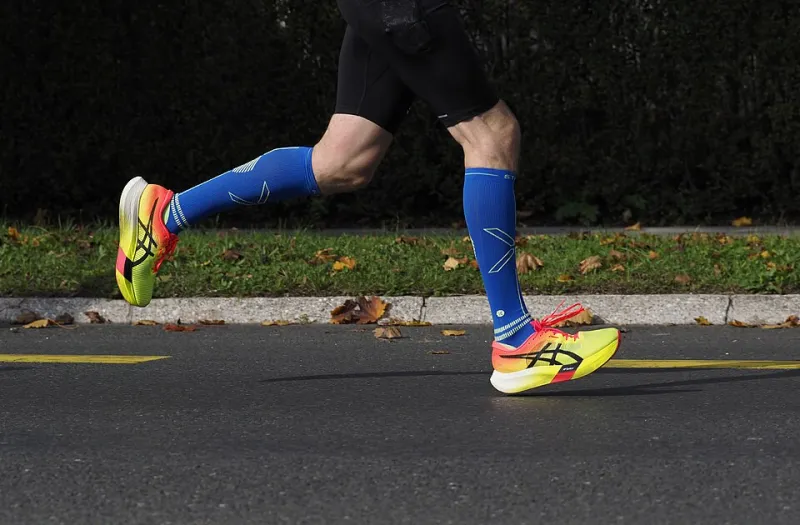
Photo: Petar Milošević, CC BY-SA 4.0, via Wikimedia Commons
It is a few minutes past nine on a Saturday morning, and somewhere in a British park a field of runners is strung out along a tarmac path. The first kilometre was sociable. The second was steady. By the fourth, conversation has stopped. Legs feel heavy, breathing is deliberate, and the finish line seems to be retreating. Many runners will describe this as 'hitting the wall' and will blame lactate, the substance they have been told is a waste product of effort. The story that cell biology tells is rather different, and considerably more elegant.
When demand outruns the supply line
Muscle contraction is powered by ATP, and at the start of a run the working fibres obtain it from several sources at once. Creatine phosphate buffers the first few seconds. Glycolysis, breaking down glucose derived from blood or from the muscle's own glycogen stores, supplies ATP rapidly but in modest yield. Oxidative phosphorylation in the mitochondria supplies far more ATP per glucose molecule, but it depends on oxygen delivery and on the capacity of the fibre to use it.
As pace increases, glycolysis is pushed harder. The enzyme lactate dehydrogenase converts pyruvate, the end product of glycolysis, into lactate, regenerating the NAD+ that glycolysis needs to keep running. This step is not a failure of metabolism. It is what allows a muscle fibre to sustain a high rate of ATP production when the mitochondrial route cannot keep pace with demand. The lactate formed is a carbon-rich molecule with considerable energy still locked inside it.
A transporter, not a drain
For a long time, lactate was treated as rubbish to be cleared. The picture changed with the recognition that muscle fibres are not identical in their metabolic wiring. Fast, glycolytic fibres tend to express the transporter MCT4, which exports lactate. Oxidative fibres, and the heart muscle in particular, express MCT1, which imports it. The two are members of the monocarboxylate transporter family, encoded by SLC16 genes, and they move lactate across the plasma membrane together with a proton.
The arrangement gives rise to what the physiologist George Brooks termed the lactate shuttle. A fibre that is producing lactate at a high rate can release it, and a neighbouring fibre with greater oxidative capacity can take it up and oxidise it for ATP. The heart, which beats continuously and prefers a mixed fuel supply, is a conspicuous consumer. During exercise, blood lactate rises from a resting concentration of roughly one to two millimoles per litre towards values that may exceed ten millimoles per litre in hard efforts, and the rise reflects production, clearance and redistribution acting together.
The liver takes part as well. Through the Cori cycle, lactate is carried in the blood to the liver, where it is converted back to glucose and returned to the circulation. The pathway costs energy, but it allows the body to recycle carbon that would otherwise be lost. Far from being a dead end, lactate sits at a junction between tissues.
The signal in the shuttle
What has changed most in recent years is the understanding that lactate is not only a fuel. It is also a signalling molecule. Lactate binds to HCAR1, formerly known as GPR81, a G-protein-coupled receptor found on adipocytes, on some immune cells, in skeletal muscle and in the brain. Activation of this receptor is linked to inhibitory G-protein signalling, which lowers cyclic AMP in the target cell. In adipose tissue, this suppresses lipolysis, suggesting that lactate can report on the state of muscular work to fat stores.
Other mechanisms are under active investigation. Lactate can influence the redox state of the cell, altering the balance of NAD+ and NADH and thereby shaping the activity of enzymes that depend on it. It has been reported to inhibit certain histone deacetylases, and to be attached to lysine residues on proteins in a process termed lactylation, which may couple metabolic state to gene expression. These findings are still being worked out, and students should read them as an emerging area rather than a settled account.
Beyond the muscle
The brain is another destination. Astrocytes, a type of glial cell, can take up glucose, convert it to lactate and pass it to neurons, which use it as a fuel. Lactate also acts on HCAR1 in the brain, where it has been linked to changes in cerebral blood flow and to aspects of neuronal excitability. The idea that exercise affects mood and cognition through many routes is not new, but lactate is increasingly included among the messengers involved.
What the wall is really made of
The discomfort at four kilometres is real, but it is not the work of a single molecule. Fatigue during prolonged or intense exercise is multifactorial: glycogen depletion in some fibres, accumulation of protons and inorganic phosphate, changes in calcium handling, rising body temperature, and signals from the brain that influence pacing. Lactate itself is not the direct cause of the burning sensation, a point that textbooks have been correcting for decades.
What lactate does reveal is a system under load, redistributing fuel between cells that have different capacities. For students, the parkrun wall is a useful teaching case precisely because it resists a tidy single-cause explanation. It shows metabolic communication in action: a signal that is also a substrate, a waste product that is also a currency, and a body that coordinates its tissues rather than leaving each to fend for itself.
By the time the runners reach the finish funnel, most of the lactate has already been cleared. The transporters and the liver and the heart have been working throughout. The lesson is not that lactate is harmless, but that it is purposeful, and that the difference between a waste product and a signal is often a matter of where the molecule travels next.